


Oral 2023, 3(3), 372-388; https://doi.org/10.3390/oral3030030 - 07 Aug 2023
Abstract
►
Show Figures
Aim: Our previous three-year randomized control trial showed that the application of MI Varnish™ (5% NaF/CPP-ACP) every 3 months reduced further caries development in 6- and 12-year-olds over a 3-year period. The purpose of this secondary analysis was to investigate whether MI Varnish™
[...] Read more.
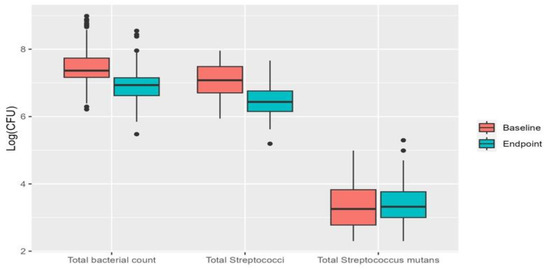
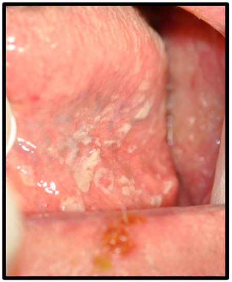
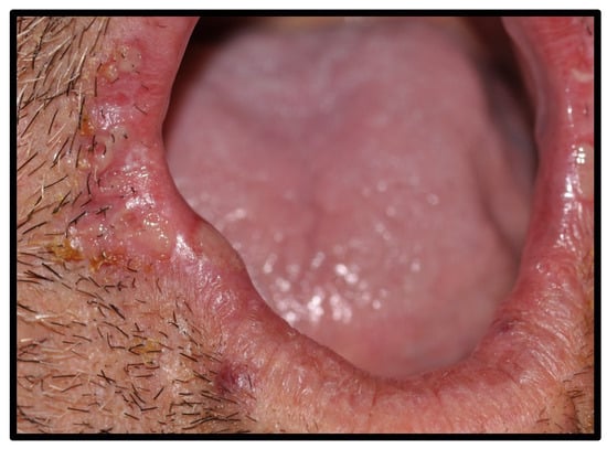
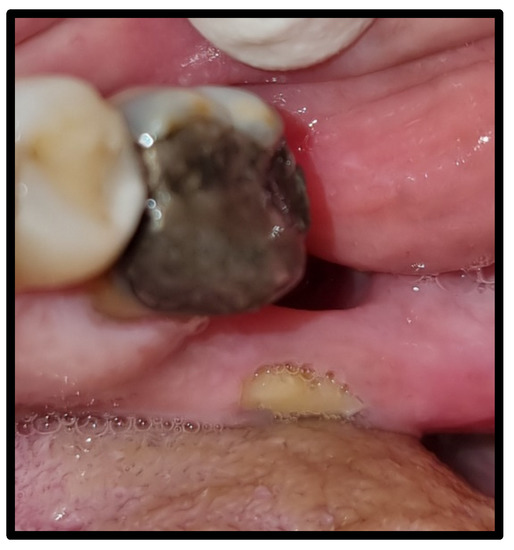
Aim: Our previous three-year randomized control trial showed that the application of MI Varnish™ (5% NaF/CPP-ACP) every 3 months reduced further caries development in 6- and 12-year-olds over a 3-year period. The purpose of this secondary analysis was to investigate whether MI Varnish™ had a differential effect on cumulative caries increment on different tooth surfaces. Methods: Group 1 (n = 48) (6-year-old children) and Group 3 (n = 47) (12-year-old children) received quarterly varnish applications, while Group 2 (n = 48) (6-year-old children) and Group 4 (n = 37) (12-year-old children) did not receive varnish applications. ICDAS caries scoring classified lesions as non-cavitated (n/c) lesions (ICDAS 1 and 2), cavitated (c) lesions (ICDAS II 3–6), non-cavitated lesions around restorations (CARn/c), and cavitated lesions around restorations (CARc). Thus, ‘decayed’ in DFS was calculated as (ICDAS 1–6 + CARn/c + CARc). The Chi-square test, Welch test (paired-t test), risk ratio test, and Pearson correlation coefficient were used for statistical analysis (α = 0.05). Results: After comparing baseline and 36-month data, in group 1, there was a significant (p < 0.01) reduction in caries in occlusal (23.11%) and proximal (21.35%) surfaces and a non-significant reduction in buccal/lingual surfaces (5.28%). In group 2, caries reduction was significant (p < 0.01) in occlusal surfaces (38.52%) but non-significant in proximal (7.78%) and buccal/lingual (7.12%) surfaces. In groups 3 and 4, significant (p < 0.001) increases in caries were observed in proximal (36.03% (group 3)/54.30% (group 4)) and buccal/lingual surfaces (51.02% (group 3)/45.98% (group 4)), and a non-significant increase was observed in occlusal surfaces (11.49% (group 3)/22.01% (group 4)). The relative risk had increased by 4% only on proximal surfaces in 6-year-olds. Conclusions: the application of MI Varnish™ every 3 months demonstrated a caries reduction effect on interproximal and occlusal surfaces among 6- and 12-year-old children. (Trial registration ISRCTN10584414).
Full article

Graphical abstract


.jpg)


