by
, , , and

Onco 2023, 3(3), 165-174; https://doi.org/10.3390/onco3030012 - 26 Jul 2023
Abstract
►
Show Figures
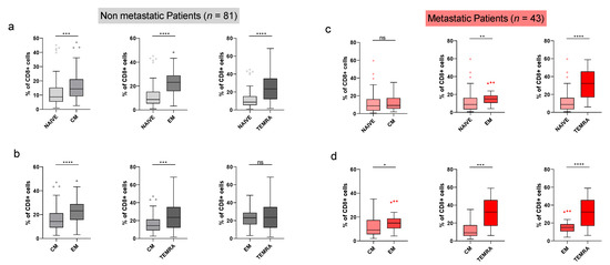
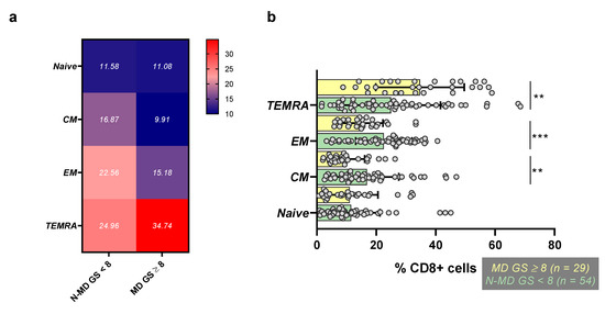
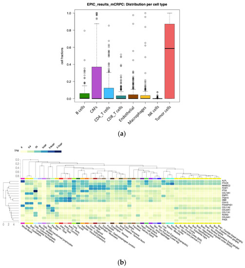
Background: Various studies have reported associations between frequencies of total peripheral blood lymphocytes and prostate cancer prognosis, but none so far has addressed the prognostic role of CD8+ T-lymphocyte subsets. Methods: A total of 43 prostate cancer patients with metastatic disease and 81
[...] Read more.
Background: Various studies have reported associations between frequencies of total peripheral blood lymphocytes and prostate cancer prognosis, but none so far has addressed the prognostic role of CD8+ T-lymphocyte subsets. Methods: A total of 43 prostate cancer patients with metastatic disease and 81 patients with non-metastatic disease were included in this study. Flow cytometry analyses were employed for determining the frequencies of peripheral CD8+ T-lymphocyte subsets. Results: Statistically significant lower levels of terminally differentiated effector (TEMRA) cells in patients with non-metastatic disease vs. patients with metastatic disease were observed. Central memory (CM) and effector memory (EM) CD8+ subsets, were found to be significantly higher in patients with non-metastatic disease vs. patients with metastatic disease. A similar profile was revealed when these CD8+ subsets were analyzed based on the patients’ Gleason scores, as well as by combined disease stage (i.e., non-metastatic vs. metastatic disease) and Gleason score. Conclusions: Peripheral blood-derived CD8+ T-lymphocyte memory subsets could function as biomarkers for the prognosis of PCa.
Full article

Figure 1


