


by
, , , , , , , and


J. Dev. Biol. 2023, 11(3), 33; https://doi.org/10.3390/jdb11030033 - 15 Jul 2023
Abstract
►
Show Figures
Receptors for advanced glycation end-products (RAGE) are multi-ligand cell surface receptors of the immunoglobin superfamily prominently expressed by lung epithelium. Previous experiments demonstrated that over-expression of RAGE by murine alveolar epithelium throughout embryonic development causes neonatal lethality coincident with significant lung hypoplasia. In
[...] Read more.
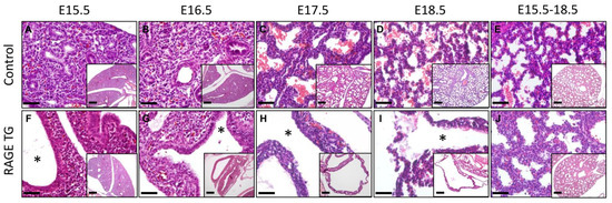
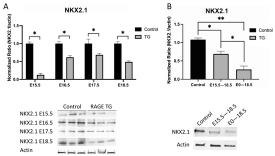
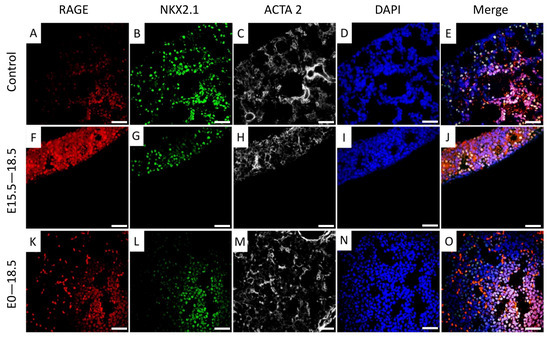
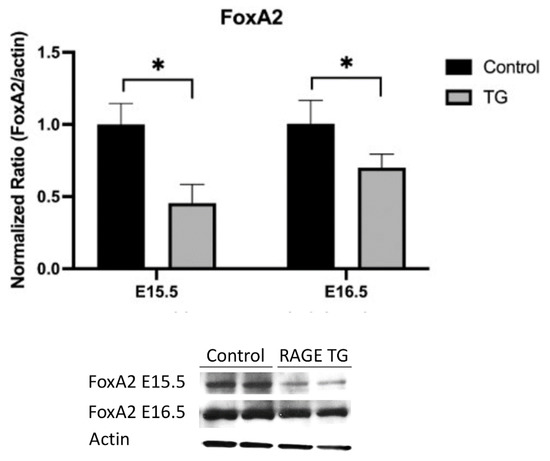
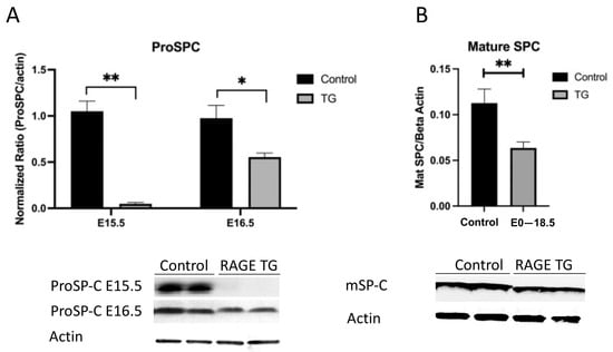
Receptors for advanced glycation end-products (RAGE) are multi-ligand cell surface receptors of the immunoglobin superfamily prominently expressed by lung epithelium. Previous experiments demonstrated that over-expression of RAGE by murine alveolar epithelium throughout embryonic development causes neonatal lethality coincident with significant lung hypoplasia. In the current study, we evaluated the expression of NKX2.1 (also referred to as TTF-1), a homeodomain-containing transcription factor critical for branching morphogenesis, in mice that differentially expressed RAGE. We also contextualized NKX2.1 expression with the abundance of FoxA2, a winged double helix DNA binding protein that influences respiratory epithelial cell differentiation and surfactant protein expression. Conditional RAGE over-expression was induced in mouse lung throughout gestation (embryonic day E0–18.5), as well as during the critical saccular period of development (E15.5–18.5), and analyses were conducted at E18.5. Histology revealed markedly less lung parenchyma beginning in the canalicular stage of lung development and continuing throughout the saccular period. We discovered consistently decreased expression of both NKX2.1 and FoxA2 in lungs from transgenic (TG) mice compared to littermate controls. We also observed diminished surfactant protein C in TG mice, suggesting possible hindered differentiation and/or proliferation of alveolar epithelial cells under the genetic control of these two critical transcription factors. These results demonstrate that RAGE must be specifically regulated during lung formation. Perturbation of epithelial cell differentiation culminating in respiratory distress and perinatal lethality may coincide with elevated RAGE expression in the lung parenchyma.
Full article
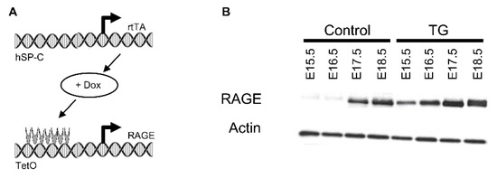
Figure 1


